Brachial Anatomy Anatomy Book
The brachial plexus is a complex intercommunicating network of nerves formed by spinal nerves C5, C6, C7, C8 and T1. The brachial plexus, frequently appears in examination questions. This guide will cover the brachial plexus and includes a summary diagram. One of the best ways to memorise the brachial plexus is by drawing it.
How to Draw the Brachial Plexus in Under 2 minutes!!! YouTube

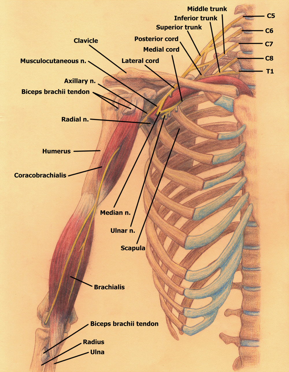
The brachial plexus is a vast network of nerves originating from the anterior rami of C5 to T1, which extends through the axilla into the shoulder, arm, and hand, providing afferent, or sensory, nerve fibers from the skin, as well as efferent, or motor, nerve fibers to the muscles. Alright, so, the brachial plexus is divided into five roots.
How to Draw the Brachial Plexus Study with an SPT

The brachial plexus is a network of nerves that originate in the spinal cord in the neck, travel down the neck (via the cervicoaxillary canal) and into the armpit. It contain the nerves that, with only a few exceptions, are responsible for sensation (sensory function) and movement (motor function) of the arms, hands, and fingers..
Brachial Plexus Injury Treatment Propel Physiotherapy
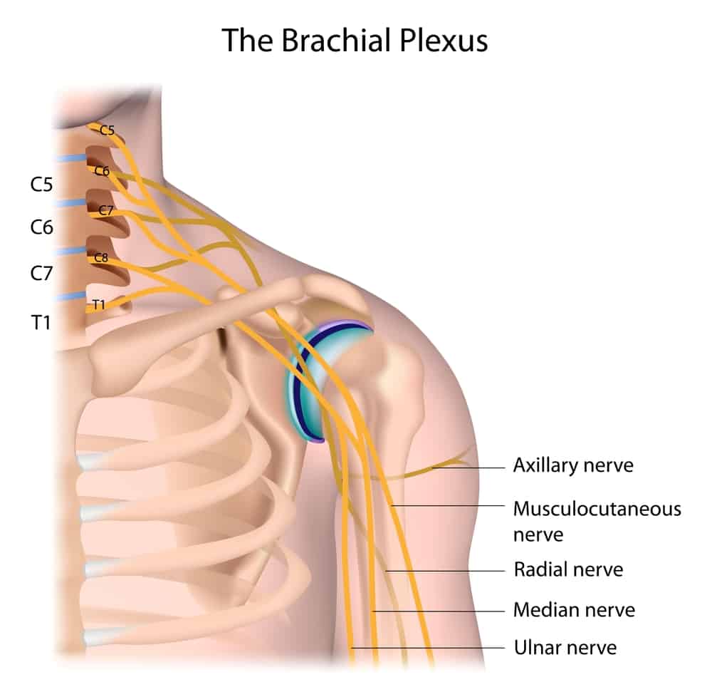
The brachial plexus is a network of nerves that originate from the spinal cord in the neck and extend down through the shoulder, arm, and hand. This complex network of nerves controls movement and sensation in the upper limb. Brachial plexus drawing is an essential skill for medical professionals, especially those focused on treating patients.
Nerve Drawings // The Brachial Plexus and its Course through the Upper
)
Check us out on Facebook for DAILY FREE REVIEW QUESTIONS and updates! (https://www.facebook.com/medschoolmadeeasy) Check out our website for TONS OF FREE REV.
Brachial Plexus Drawing AlyssaafeHawkins

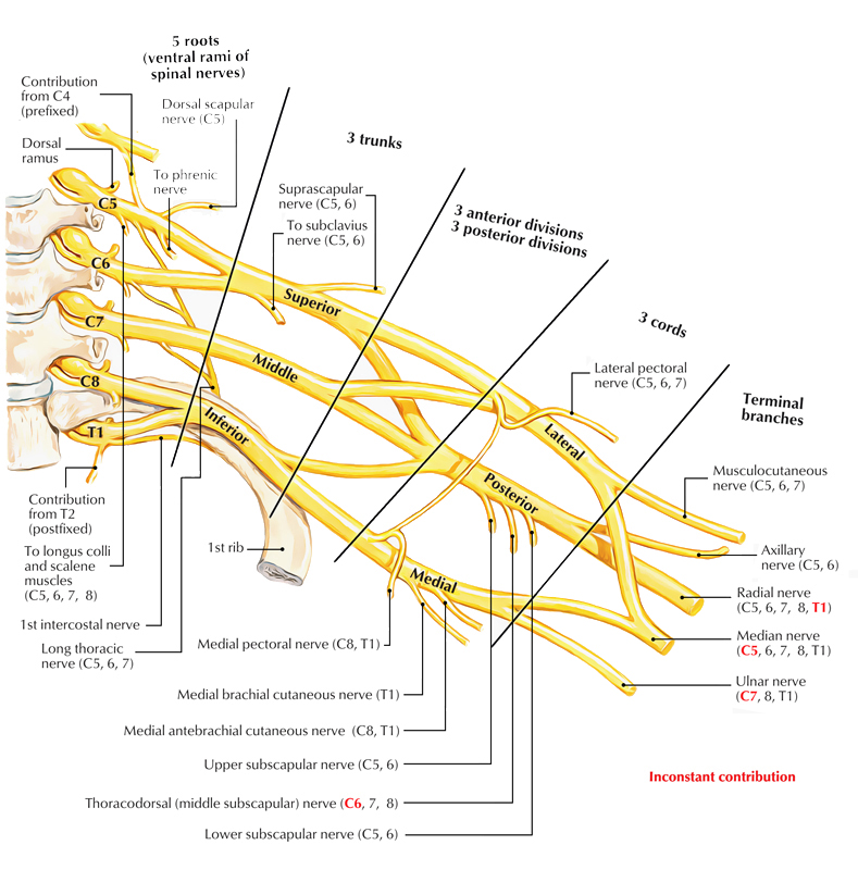
The brachial plexus is a network of nerve fibres that supplies the skin and musculature of the upper limb. It begins in the root of the neck, passes through the axilla, and runs through the entire upper extremity. The plexus is formed by the anterior rami (divisions) of cervical spinal nerves C5, C6, C7 and C8, and the first thoracic spinal nerve, T1.
Brachial Plexus Art as Applied to Medicine

The most serious brachial plexus injury occurs when the nerve root is torn from the spinal cord. Signs and symptoms of more-severe injuries can include: Weakness or inability to use certain muscles in the hand, arm or shoulder. Complete lack of movement and feeling in the arm, including the shoulder and hand. Severe pain.
Anatomy of the brachial plexus. Download Scientific Diagram

The brachial plexus is a network of nerves that gives rise to all the motor and sensory nerves of the upper extremity.This plexus arises from the anterior rami of spinal nerves C5-T1 that undergo several mergers and splits into trunks and divisions, until they finally give rise to their terminal branches.These terminal branches are responsible for motor and sensory innervation of the upper.
The Brachial Plexus Sections Branches TeachMeAnatomy

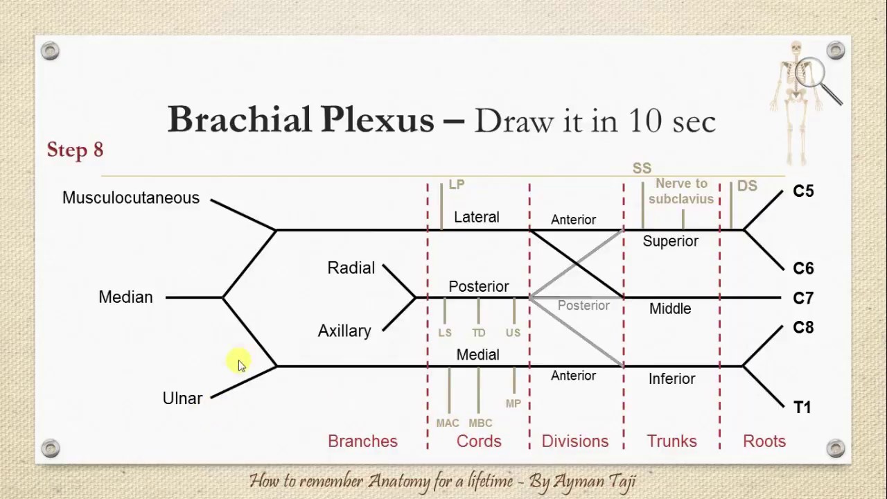
How to draw a brachial plexus in just 1 minute or 60 seconds ?. This educational video brought to you by Medchrome perfectly answers this question.. Step by step instructions for drawing a brachial plexus that looks real: 1. Write the Root values C5, C6, C7, C8 and T1 leaving almost equal space between the 2 consecutive points.
How to Draw the Brachial Plexus Physical therapy student, Physical

This is an instructional video on how to diagrammatically draw the brachial plexus along with explanations. The diagram takes only a minute to draw but the v.
Brachial Plexus Anatomy and Clinical Correlation
An easy way to draw the basic components of the brachial plexus. On paper, it's pretty easy to draw the brachial plexus using this method in around 10 secon.
Branching of the Brachial Plexus (anterior view) by Blique on DeviantArt
Drawing Brachial Plexus There are several nerve plexuses in our body. The four main nerve plexuses are the cervical plexus, brachial plexus, lumbar plexus, and sacral plexus. The choroid plexus in the brain is a part of the central nervous system which consists of ventricles, capillaries, and ependymal cells.
Upper Limb Nerve Lesions (Part 1 The Brachial Plexus)
In this video, we will show you a fun way to draw and memorize the main structure of the brachial plexus. To learn all the details about the anatomy of this.
Brachial Plexus Anatomy, Location, Function, Injury and FAQs
Learn how you can remember and draw the brachial plexus in just 10 seconds!~~~~~This video is a part of my anatomy co.
Here is a "CheatSheet" for the Brachial Plexus I Made for any New
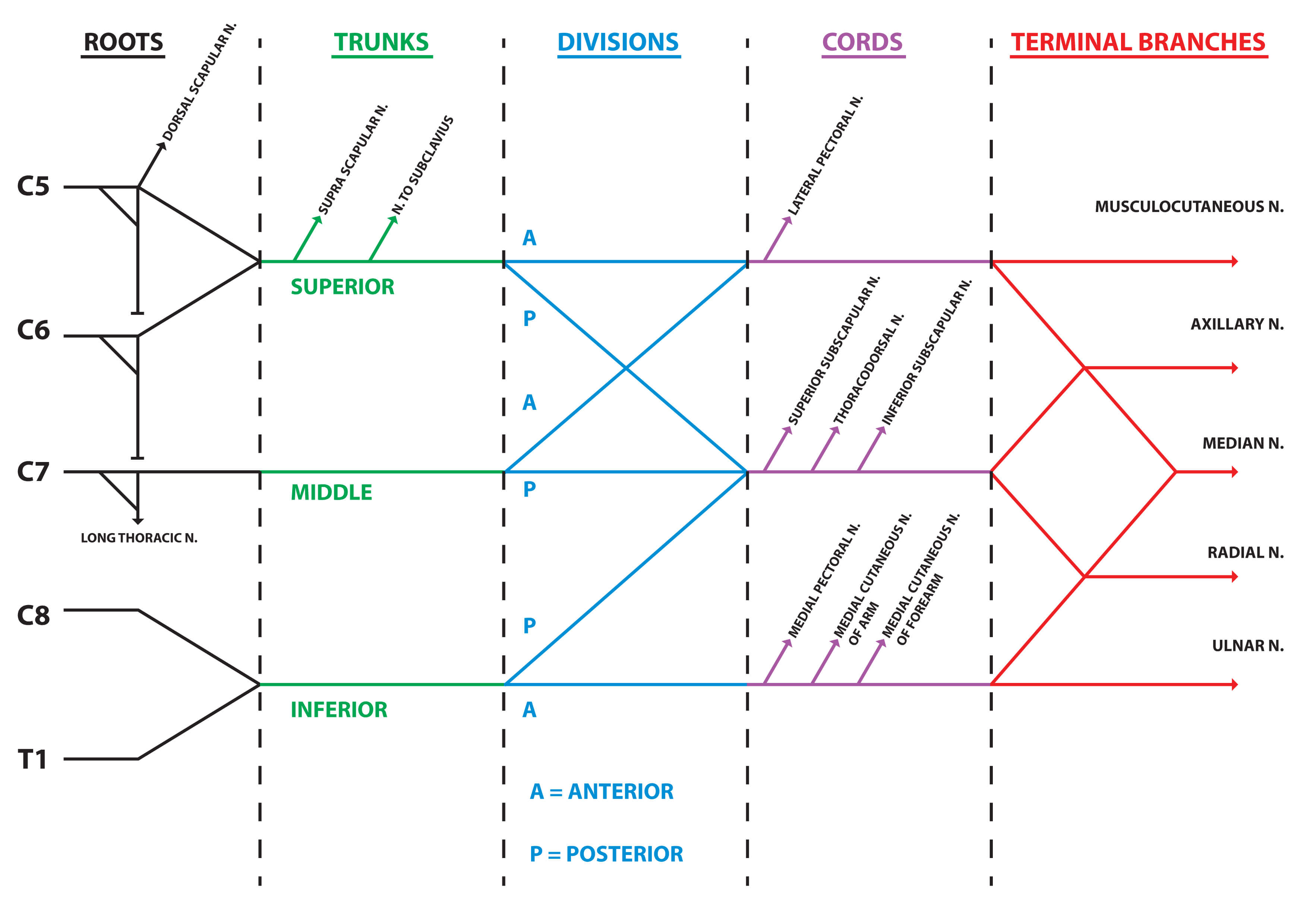
Objectives: Be able to draw the brachial plexus in your sleep. Learn to identify the roots, trunks, divisions, cords, and individual. branches by using the interactive images. Link the various terminal branches with their specific origins and spinal root components. Equate the various major plexus related traumas with their specific site of injury.
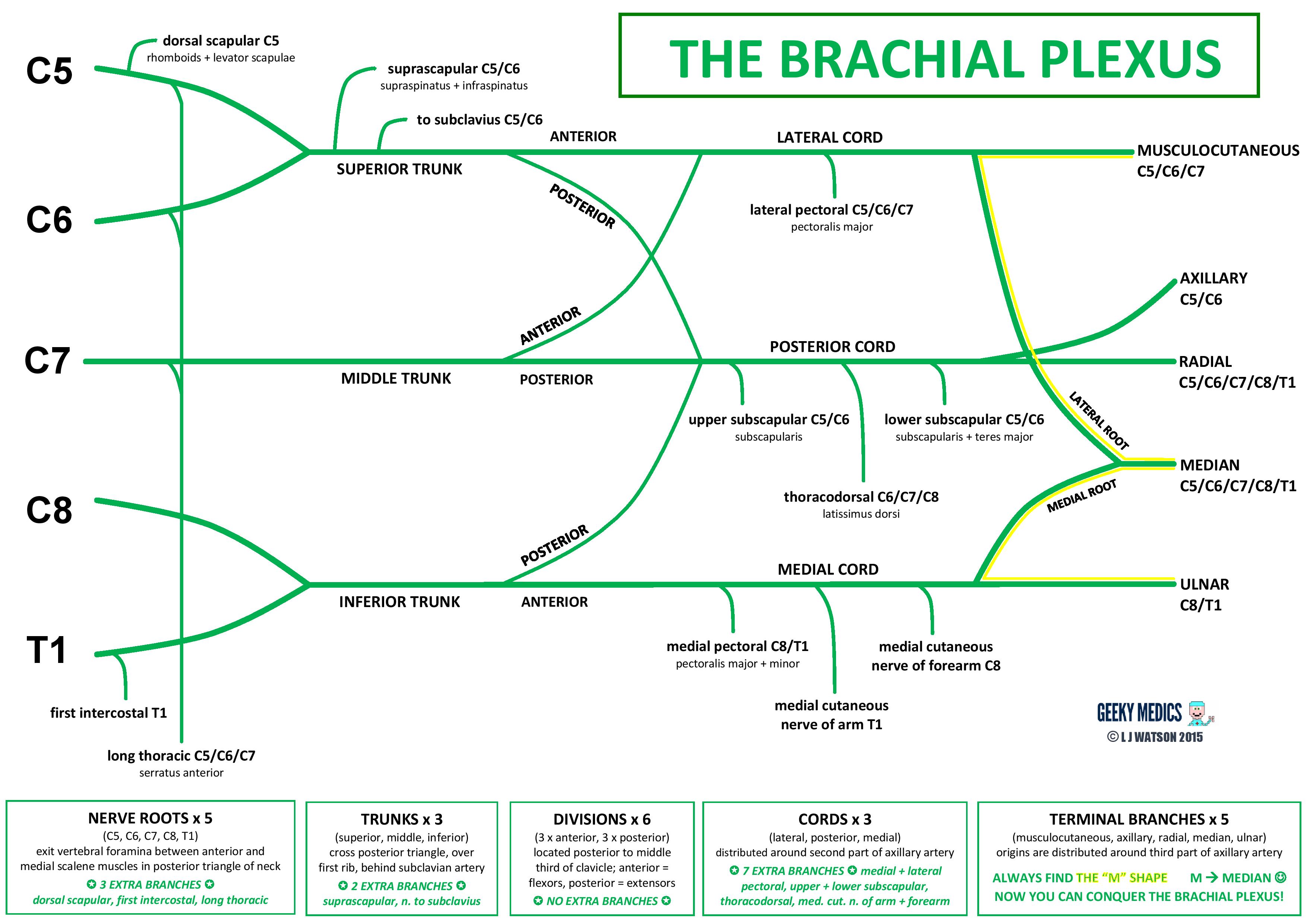
Brachial Plexus Anatomy Roots Trunks Cords Geeky Medics
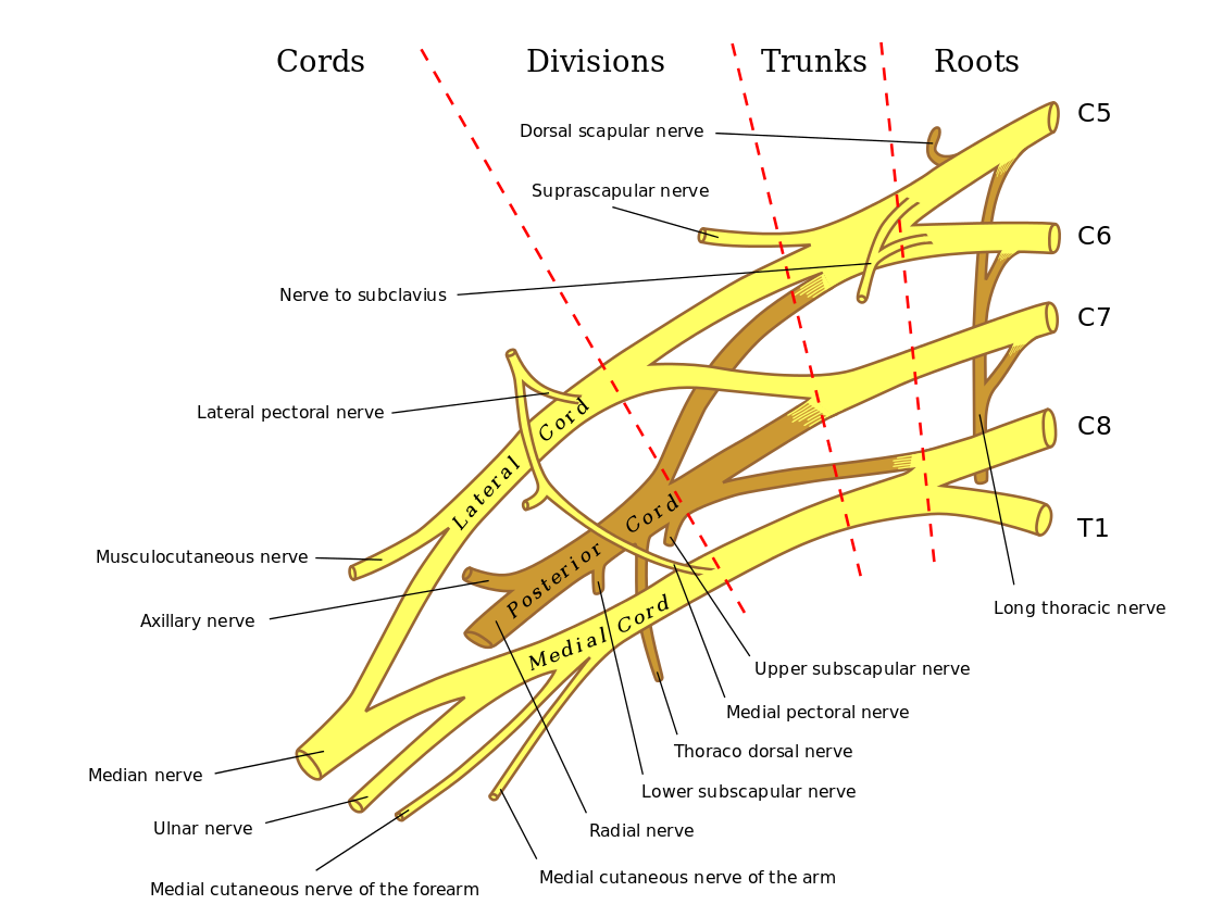
Normal sagittal appearance of the brachial plexus. Drawing (a) illustrates the roots, trunks, divisions, cords, and terminal branches of the brachial plexus, which grossly appear on CT images (b) but are much better seen on MR images (c) obtained with T2-weighted fat-suppressed (top row in c) and T1-weighted (bottom row in c) sequences.